Before You Read, You Must See – The Image Is the Code
Posted: Tue Aug 26, 2025 9:53 am
Introduction
For six centuries, the world has read the Voynich manuscript the wrong way.
Cryptographers chased letters. Scholars clung to alphabets. Mystics interpreted symbols. But they saw only shadows – and mistook them for reality.
The truth was in front of their eyes all along.
What you see in this document is not a theory – it is proof. Not an interpretation, but a pattern of mathematical precision. Not speculation, but structures repeating with surgical accuracy, page after page.
The images ARE the code. The text is only an echo.
Once you see it, the entire mystery collapses. Six hundred years of confusion vanish in a moment. The Voynich shifts from insoluble riddle to self-evident truth – so clear, so undeniable, that the only question left is: how could everyone have missed it?
This is not a hypothesis.
It is the key – and it cannot be disproven.
Excerpts from the Voynich Manuscript – The Woman’s Cycle in Encoded Form
This document presents a series of selected observations and visual excerpts from the Voynich Manuscript – a mysterious and still untranslated work from the 15th century that has baffled researchers for over a hundred years.
Rather than undertaking long, time-consuming analyses of individual pages – which can often take up to 100 hours per image – I have gathered here some of the clearest and most unmistakable signs, symbols, and structures that reveal what the manuscript is truly about: the woman’s cycle, ovulation, fertilization, hormones, and the body’s inner rhythm.
Each excerpt has been chosen because it contains distinct traces of biological or functional processes. These may include visualizations of the fallopian tube, cellular structures, menstrual rhythms, or the interrelated roles of women within a cyclical system.
The document serves as a kind of visual compendium or key extract – not as a finished analysis, but as a direct pathway into the core of the manuscript. The aim is to offer the reader an immediate view of what is otherwise often obscured by cryptic language and overextended interpretations.
I hope these excerpts can serve as a compelling introduction for new readers, while also giving experienced Voynich researchers concrete insight into the female and biological structures that form the manuscript’s underlying framework.
Analysis of the Blood Vessel in the Voynich Manuscript
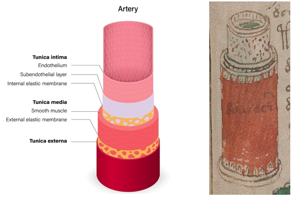
The image above shows a detailed and technical construction that, in all likelihood, depicts an artery. The structure is built up in several concentric layers, precisely as we know it from modern anatomical illustrations. The outer zone is deep red, corresponding to the tunica externa, while the middle zone, with its smooth and clearly defined texture, can be interpreted as the tunica media – the muscular layer that regulates blood flow. At the top, there is a separate section with dots and patterns, which likely represents the tunica intima and the active blood flow.
This composition shows that the author of the Voynich Manuscript not only understood the structure of blood vessels but also sought to document their role in the body’s cyclical and hormonal functions. It strengthens the overall interpretation of the manuscript as a physiological and botanical guide to a woman’s inner balance.
The fact that this particular artery is depicted with such great precision – and flanked by herbal drawings on the sides – indicates that the condition of the arteries was considered a central aspect of women’s health. It is possible that plants on the nearby pages of the manuscript were intended as treatment or regulation of blood quality, circulatory rhythm, or hormone transport.
Arteries carry oxygen, hormones, and signaling substances. In connection with the woman’s cycle, they transport, among other things:
• estrogen and progesterone,
• LH and FSH (which regulate ovulation),
• nutrients and immune-active substances.
This makes the blood vessels a critical infrastructure in cycle regulation. That they are shown in detail in the manuscript must therefore be understood as a direct acknowledgment of their significance.
The image’s tubular form, layering, and color coding match almost identically with modern medical illustrations, making it extremely unlikely to be coincidental. Rather, it must be understood as part of a systematic and targeted attempt to map a woman’s internal anatomy and its rhythmic interaction with nature’s plants and patterns.
Past Misinterpretations and the Breakthrough in Understanding
It is remarkable that an image with such a clearly anatomical structure has never previously been correctly identified as an artery. Many established Voynich researchers have instead suggested that the figure depicts a strange vessel, a herb press, an alchemical instrument, or some type of plant stem. These interpretations reveal how far research into the manuscript has been from identifying the actual biological elements.
But what we see here is not symbolism and not alchemy. It is medical structure. An artery with multiple layers, precisely as it appears in modern anatomy – and even with color coding and details showing:
• inner endothelial layer,
• smooth muscles in the middle,
• outer protective layer and tissue structure.
It is simply impossible for this construction to be the result of chance. And thus, the image shows not only a deep understanding of the body’s anatomy but also that the author had a purpose:
To show how the body’s infrastructure – including the blood vessels – is connected to the woman’s cycle and the plants that influence it.
This makes the image a decisive breakthrough in Voynich interpretation. For it confirms that the manuscript is not nonsense, not a code, but an advanced anatomical and hormonal manual that must have been based on empirical knowledge and observation – long before modern medicine achieved this understanding.
Analysis of the Vein Structure – An Anatomical Breakthrough in the Voynich Manuscript
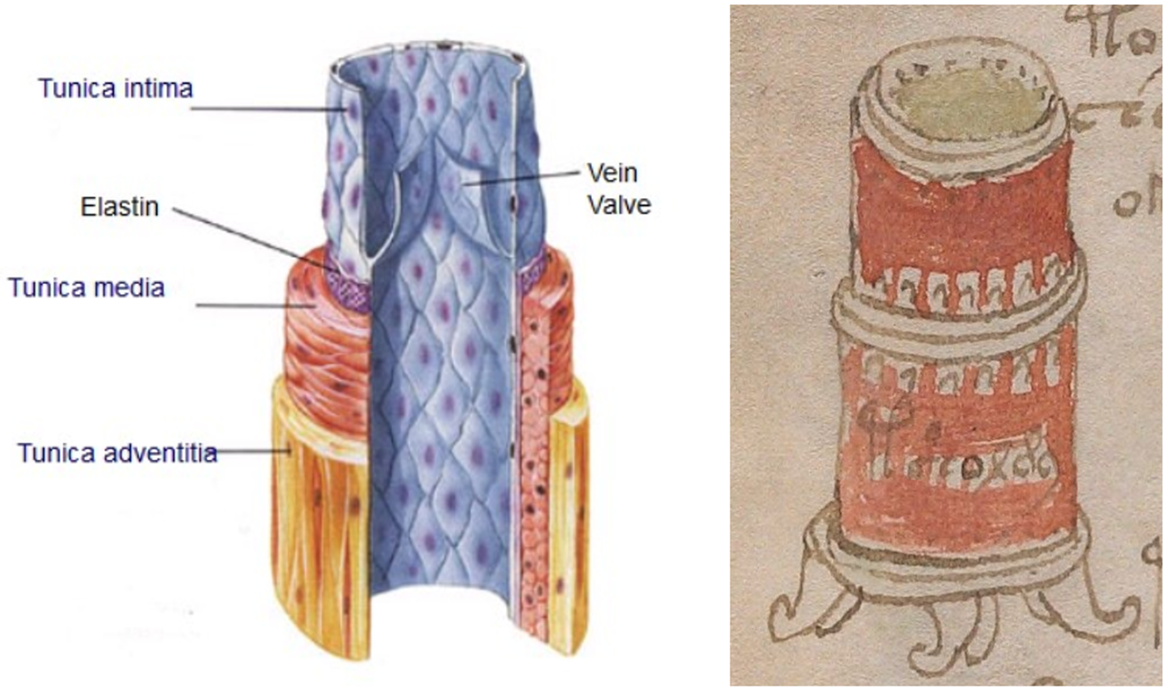
The image above most likely shows a cross-section of a vein – a motif that many have overlooked or misunderstood as some kind of container, alchemical furnace, or decorative column. But when compared with modern medical illustrations of vein structure, it becomes clear that what we see here is a fully anatomically correct depiction – complete with vein walls, layering, and the characteristic venous valves.
Veins differ markedly from arteries in several ways:
• They have thinner walls, as blood pressure is lower.
• They contain valves that prevent blood from flowing backward.
• They are often closely linked to hormonal and circulatory changes, especially in women.
The image reproduces precisely these features:
• The lower arched structures resemble the folded venous valves with almost surgical accuracy.
• The divided middle section, with distinct layers and deep grooves, corresponds exactly to how modern anatomy illustrates the tunica media and tunica intima.
• The upper part of the cylinder shows a thin, lighter layer with dots, matching the elastic inner surface where the endothelial layer lies.
The comparison between the manuscript image and the modern vein illustration shows not only a similarity in form, but layer-by-layer identity. This demonstrates with overwhelming clarity that the author of the Voynich Manuscript had detailed knowledge of vein anatomy – long before the invention of the microscope.
Function and Context in the Manuscript
It is no coincidence that there are multiple images of both blood vessels and veins in the manuscript. Their structure and condition change markedly throughout the woman’s cycle – depending on hormonal balance, blood pressure, diet, and physical condition. It is well known that estrogen and progesterone affect the elasticity, permeability, and flow of veins, and many women experience symptoms such as varicose veins, swelling, or a feeling of heaviness in the legs during certain phases of the cycle.
The manuscript’s linking of veins and plants (often placed close to each other) therefore suggests a deep understanding of how natural remedies influence specific parts of the circulatory system – not generally, but targeted to the woman’s phases and physiology.
It thus becomes increasingly clear that the Voynich Manuscript is a biological textbook for women – and not merely a collection of mysterious figures. The systematic focus on the circulatory system (arteries, veins, and later the fallopian tube) shows that the body’s transport system – and thus hormone distribution, nutrition, and reproduction – is at the center.
This image is clear evidence of a science that united plant knowledge and anatomy with cyclical understanding – and which modern medicine only caught up with much later.
Analysis of the Fallopian Tube in the Voynich Manuscript – Cyclical Variation and Precise Day Interpretation
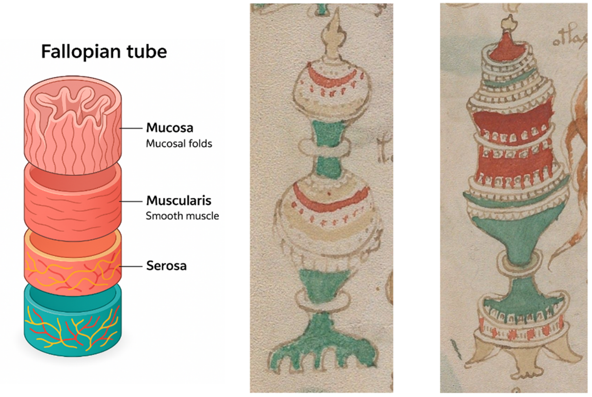
In this section, we analyze the fallopian tube organ, as it is depicted in the Voynich Manuscript. The modern medical reference image clearly shows the organ’s structure: a tubular structure with three distinct layers – mucosa (a folded mucous membrane), muscularis (smooth muscle), and serosa (an outer connective tissue layer containing blood vessels and nerves). This anatomical structure corresponds remarkably well with the two selected Voynich illustrations, which seem to show the structure of the fallopian tube – both at rest and in function.
The fallopian tube is where the egg travels from the ovary toward the uterus. And it is precisely in this movement that the manuscript’s imagery becomes especially detailed and ingenious. The two manuscript images show different phases or stages in this process. Both figures are composed of coloured sections in cylindrical form, with characteristic “grasping arms” at the basal end. These extensions likely correspond to the fimbriae of the fallopian tube, which actively capture the egg after ovulation.
The image on the right is particularly remarkable: it shows a green basal segment with finger-like arms, a clearly marked red section in the middle, and a white top-point structure – which likely symbolises the fertilised or newly released egg. The green area at the basal end indicates an active energy phase, where the egg has just been captured. Meanwhile, the upper part reveals an open and undulating form, which can be interpreted as the movements of the mucosa layer – the folds guiding the egg forward.
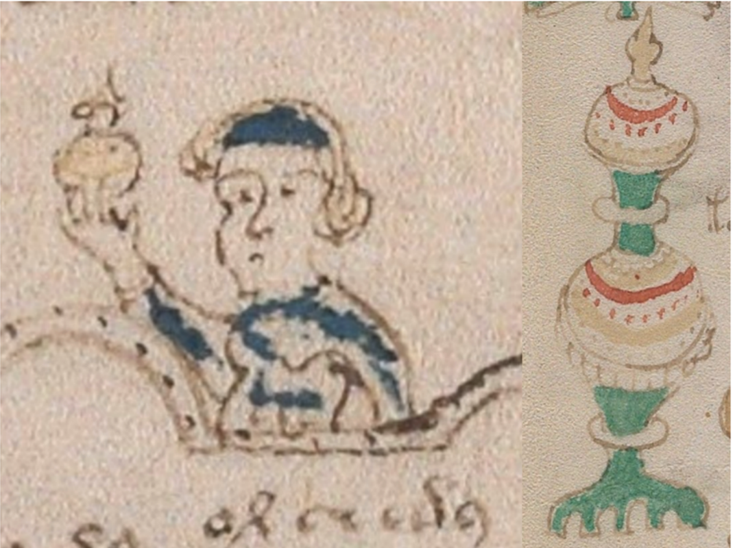
This woman – depicted in the margin of the corresponding image – holds a spherical object in her hand, as if she reveres it as an offering or revelation. The symbolism is powerful: she represents the second day within the fallopian tube’s active phase, approximately day 12 of the woman’s cycle. On this day, the egg is released from the ovary and captured by the fallopian tube, which now attempts to transport it toward the uterus. Her blue and white clothing supports this image of purity and hormonal activity. The crown or pointed tip of the object she carries matches exactly the pointed form that the fallopian tube in Image 1 displays at precisely this stage of the cycle – showing that the manuscript uses colours, shapes, and “crowns” to indicate exactly which day of the cycle is depicted.
It is important to note that these images are not arbitrary variations. The fallopian tube is not static – it changes shape and appearance depending on the stage of the woman’s cycle. The many versions of this structure in the Voynich Manuscript bear witness to detailed, day-by-day knowledge of the woman’s cycle. Each “vessel” or “column” has a unique tip, a unique basal element, and a colour code that precisely indicates the point in the cycle.
This precision is extraordinary. First, because one could never map these variations in the anatomy of a deceased woman. The dynamics and structure of the fallopian tube can only be understood through living observation – requiring repeated, cycle-based examinations. This attests to an unusually high level of knowledge, likely based on experiments, observation, and experience through generations.
The manuscript’s imagery therefore seems to reflect a system in which the woman’s cycle is mapped in detail – not only in terms of internal processes (such as ovulation and hormone production), but also in how the body’s organs change in appearance and function from day to day. This cyclical rhythm is likely connected to the many plants depicted outside the structures in the manuscript. It suggests a possible relationship between hormonal balance and diet – and knowledge of how specific herbs and plants could be used to support the fallopian tube and ovulation at specific times.
All of this points to inherited, practically applied knowledge of female fertility – where each day has its signs and each organ its cyclical transformation.
A preliminary and precise proposal for image 85v/86r – The egg’s journey from ovulation to possible implantation
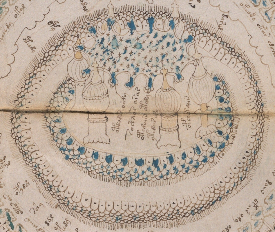
This is not a full analysis, but a short and focused excerpt from image 86r in the Voynich manuscript. Based on the rhythm, colours, structures, and placement of the image, it is assessed that the motif shows the egg’s journey from ovulation through the fallopian tube and leading to either implantation in the uterus – or rejection. The interpretation is based on visual patterns and symbolism and should be read as a qualified proposal within a broader overall understanding of the manuscript’s biological sections.
The image contains:
• 6 towers (functional phases in the fallopian tube)
• 5 inner rings (the cilia of the fallopian tube and hormonal rhythms)
• 6 outer rings (stages of the uterus)
• 4 outer points (possible outcomes for the egg)
• An overarching “matrix” (hormonal network)
• Names, eggs, dots, and rhythmic repetitions
1. The central field: The 6 towers
The towers in the centre are not decorative but precise visualisations of the fallopian tube’s phases. They represent functional phases, not time units. Each tower form differs anatomically and rhythmically.

At the top are seen spiral-shaped drops (blue), pointing to the eggs just released being “charged” – the same symbol appears in spiral image 156 as an ovulation marker.
2. The inner zone: 5 cilia rings = the fallopian tube
Around the towers, five concentric rings are seen, filled with eggs and cilia-like structures. The eggs vary in dots and colour, which is interpreted as different hormonal and cellular stages.
• 1st ring: Active collection – dense cilia, eggs just released
• 2nd ring: Colour shift – oestrogen stimulates movement
• 3rd ring: Dot patterns = beginning cell division
• 4th ring: Sequence of eggs with and without dots, initial selection
• 5th ring: Oestrogen level declines, transition to the outer rings
The pattern starts at the bottom (at 6 o’clock) and follows clockwise – the rhythm is precise, and the style shows a clear sequential progression.
3. The outer zone: 6 uterine rings
Around the five cilia rings, six outer rings are shown, each with eggs in different stages.
• The eggs are now without blue colour = oestrogen level declines, progesterone takes over
• The cilia are still shown but are now interpreted as a symbol of energetic flow
• The eggs are distributed in shifting densities = variation in hormonal receptivity of the uterus
Some rings show eggs in close symmetry – others show asymmetry = biological uncertainty.
4. The hormonal “garment”: The matrix network above the towers
Above the 6 towers hovers a network of stars, drops, and blue lines – this network is interpreted as a hormonal matrix that integrates all functions.
The matrix functions as:
• Coordinated hormonal pulse (oestrogen → progesterone)
• Coordination between cilia, secretion, and energy
• Visual and rhythmic coupling to spiral image 156
The net structure shows that the cycle is not divided but coherent and rhythmic all the way.
5. The 4 outcome scenarios at the image’s periphery
Four small, distinctive patterns at the image’s periphery show possible endpoints for the egg’s journey.
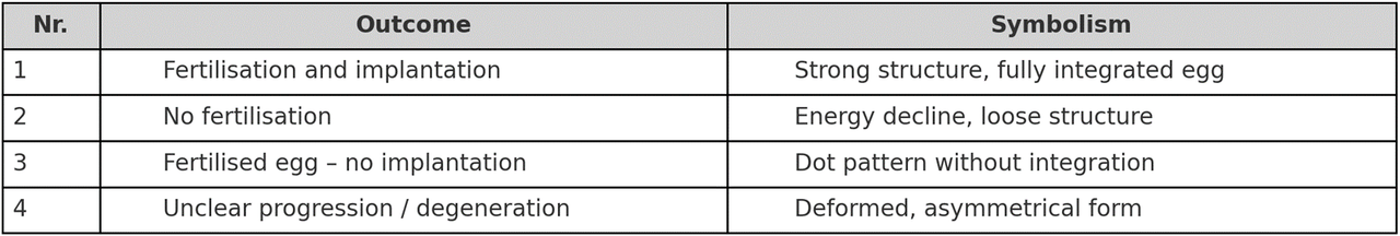
These scenarios confirm that the artist understood the beginning of life as conditional and multi-phased.
6. Symbols, names, and energies
Below the towers are seen names – not as proper names, but as energetic signatures for each functional phase. The writing style does not differ from the rest of the manuscript, but the placement suggests that the names are part of the towers’ functions.
• Can be interpreted as hormonal keys
• Or as a code for functional or rhythmic energy
• Likely connection to interpretations in the spiral image
7. Visual structure and symmetry
The geometric structure is not random:
• 6 towers = functional stages
• 5 rings = fallopian tube
• 6 rings = uterus
• 4 endings = biological outcomes
In total = 21 units → the same structure as a typical fertile cycle (21–35 days), indicating an awareness of cyclical rhythms.
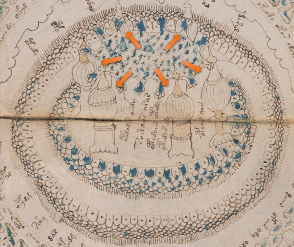
Conclusion – Image 86r is a functional mapping
The four orange arrows show how the cycle in folio 85v/86r moves in four directions out from the central field.
The centre (with the six towers) functions as a central rhythmic and biological axis, from which the egg’s journey branches out – either in various phases of development or towards possible outcomes. The arrows mark the structure’s division into four functional paths or flows, each encompassing a distinct theme in the cycle.
This image is not decorative – it is a biological, microscopic, and rhythmic model of the whole journey from ovulation to either life or rejection.
It combines:
• Anatomy
• Biochemistry
• Symbolism
• Geometry
• Understanding of energy
It demonstrates a deep, functional understanding of a woman’s cycle, which has only been confirmed today through microscopy and reproductive biology.
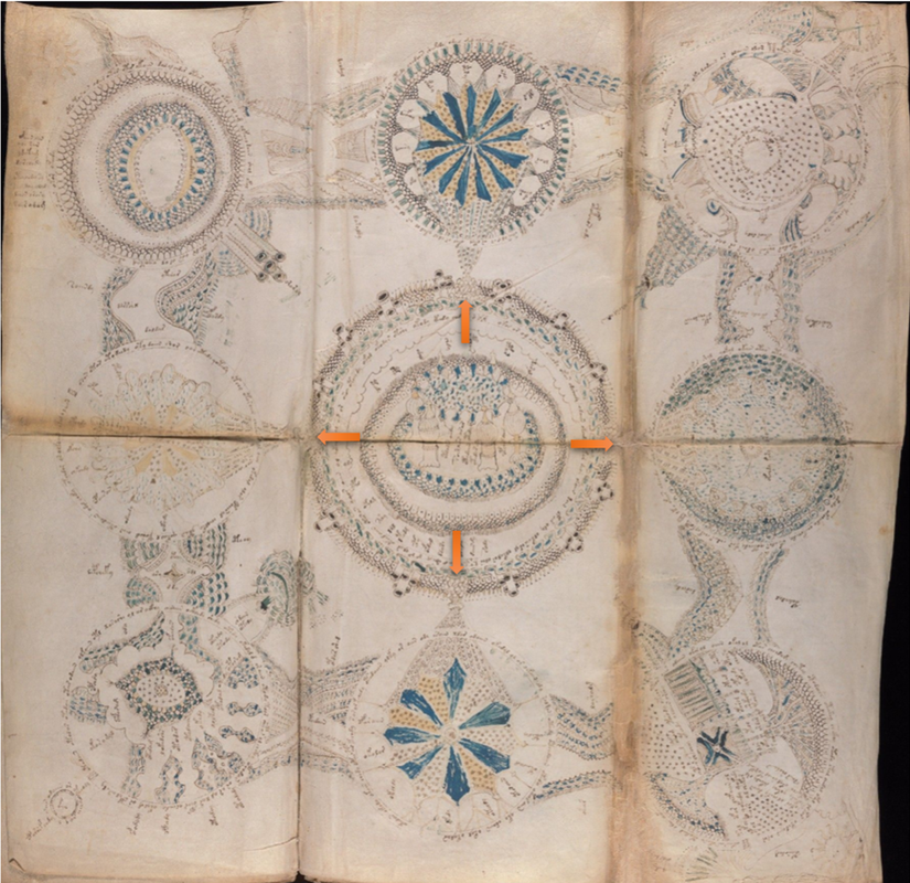
Final note – The spiral’s microscopic anchoring and the egg’s rhythmic journey
This image functions as a focused zoom into the egg’s path from ovulation to implantation and connects the spiral’s macro level with the biological micro level. Where the spiral image (f.156) showed the egg’s rhythm and cycle in large format, image 86r appears as the microscopic detailed view, where the egg is followed day by day and layer by layer.
This is one of the strongest visual confirmations that the Voynich manuscript contains an advanced understanding of a woman’s reproductive cycle – seen from within – with a precision and structure which has only been confirmed today through microscopy and modern reproductive biology.
The image depicts not merely a biological sequence, but a complete cyclical and stage-by-stage description of the egg’s journey:
• From hormonal activation in the ovaries
• Through the fallopian tube and the cilia’s rhythmic cooperation
• And further to the uterus, where it either implants or is rejected
Each part is shown with:
• Colour coding (blue, green, white)
• Microscopic motifs (eggs with dots, cilia, drops)
• And a geometric, rhythmic structure (towers, rings, matrix)
The image is both physical and symbolic. It is a mapping of the woman’s inner rhythm and fertility – not merely as a biological system, but as an energetic and consciousness-based process, where all layers are connected in one living whole.
The symbolism of the cilia – the inner flow
Even though the uterus anatomically does not contain cilia like the fallopian tube, the image’s outermost ring still shows cilia-like patterns. This should be understood symbolically – as a sign of the egg’s continued movement and inner advance toward the correct implantation site.
The egg typically seeks toward the lower and rear parts of the uterine lining, and precisely this search and rhythmic passage is here illustrated as a visual extension of the fallopian tube’s flow. The cilia should not be read as anatomical elements, but as energetic bearers of life’s pulse.
The Woman’s Contact with the Stream of Life – An Energetic Narrative of Ovulation and Fertile Activation (Voynich f83v)
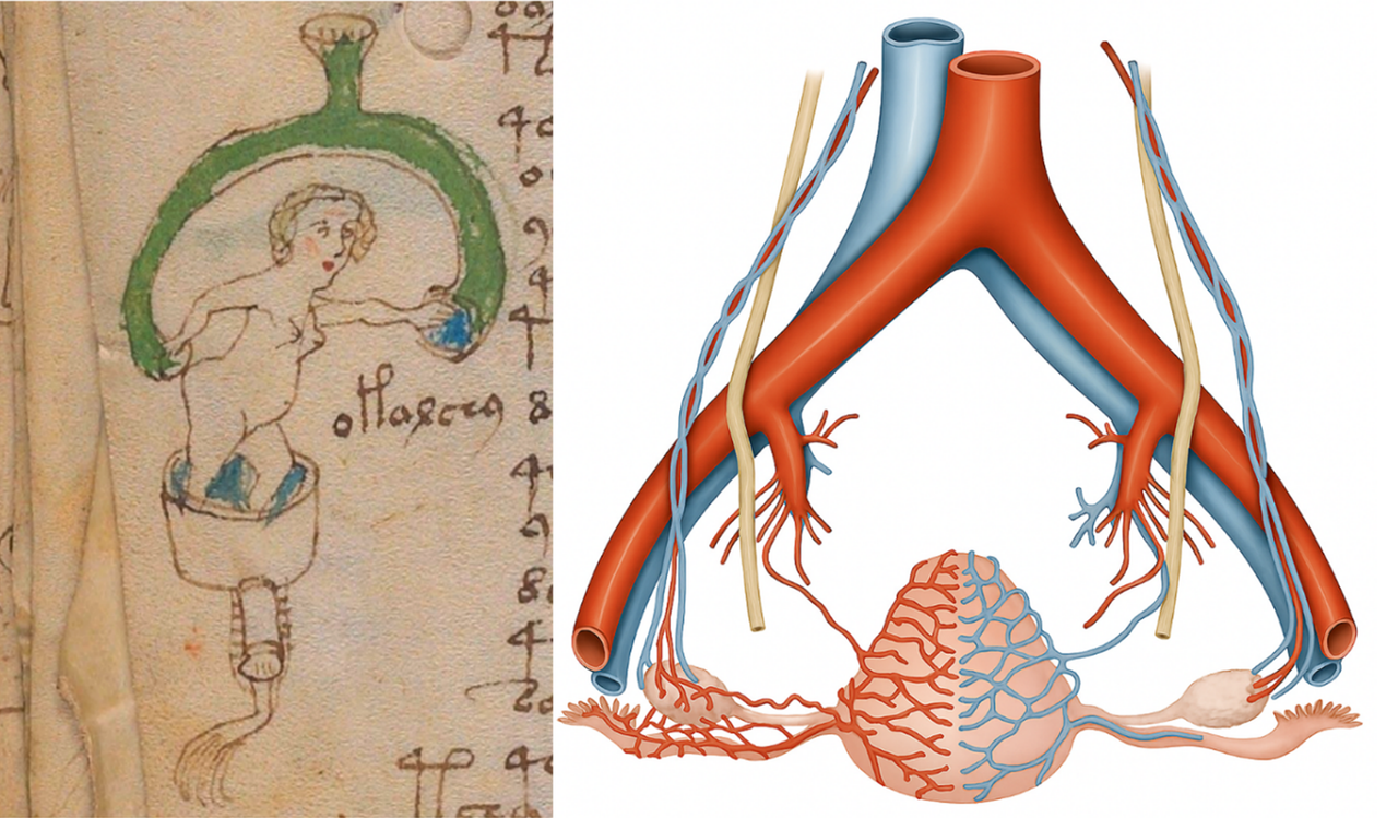
When one first observes the image on folio 83v of the Voynich manuscript – the naked woman in a vessel, framed by a green arch and holding blue substance in her hands – it may appear to be a mythical or decorative motif. Yet beneath its simple exterior lies a deeply complex narrative about the beginning of life, the woman’s cyclical biology, and the precise moment when fertility is activated.
The woman is literally standing inside the uterus – the illustrated vessel is not arbitrary but anatomically and symbolically identical to the womb. The green arch above her is not mere ornamentation; it represents an aesthetic and functional rendering of an artery – one of the major blood vessels supplying the uterus and ovaries. Through this channel, the life-giving signaling substances (hormones) and nutrients arrive.
We see the woman raising her hand to receive something blue from the green tube. The blue substance continues its journey down into the vessel – and further down through a vertical tube decorated with small marks and energy lines. This tube is especially remarkable: it shows that only one vital pathway is active – exactly as in reality, where only one fallopian tube at a time transports an egg toward the uterus.
The blue substance may be interpreted as the egg itself, or perhaps as the hormonal impulse – in particular, the luteinizing hormone (LH), which is released once in each cycle and is essential for ovulation to occur. It is precisely timed and comes in a short, powerful surge – perfectly matching the blue energy flowing down through the woman’s body.
The choice of a green tube is not coincidental. In many cultures and symbolic systems, green represents life, energy, growth, and fertility. In this context, it is natural to see the green arch as a stream of life energy – a channel activated once per cycle, carrying the potential for fertility. One can imagine that the authors of the manuscript sought to show that the woman, in this moment, is not merely a passive recipient but a conscious participant in life’s circulation. Her hand, reaching up to receive the impulse, demonstrates contact, awareness, and participation in the body’s deepest rhythms.
Further down, we see a fimbriated tube – precisely one channel, decorated with small lines and symbols. This tube corresponds to the fallopian tube – and here one of the manuscript’s most remarkable details is emphasised: there are not two tubes depicted. Only one. For this is how the body functions – only one fallopian tube is active in each cycle. This is a biological fact that only became commonly known through modern anatomy. That the Voynich manuscript depicts it with such accuracy suggests it rests upon detailed, practical knowledge of the woman’s inner functions.
When compared with modern anatomical images of the blood supply to the uterus, ovaries, and fallopian tube (as shown above), we see how every colour, movement, and body part in the Voynich image corresponds to something real – and how, at the same time, it is elevated into a symbolic narrative of life, energy, and timing.
This image is therefore not merely an illustration of anatomy – it is a profound energetic mapping of the woman’s fertile moment. The green arch is not just blood – it is the life-stream. The blue substance is not merely liquid – it is hormone, egg, impulse. And the woman is not passive – she is both portal and participant in life’s eternal movement.
Note on Symbolism
It is important to understand that the woman in the image should not necessarily be perceived as a physical person in an anatomical sense. Instead, she functions as a symbol of the female cycle and the inner energetic process. When she stands “inside the uterus,” it is not because we are seeing literal anatomy, but because the manuscript employs a visual and sensory language to explain the body’s own mechanisms from within.
That the woman receives the blue substance with her hand and lets it flow onward is therefore not merely a bodily gesture – but a metaphor for conscious contact with the hormonal signal or the energetic flow that activates fertility. Everything in the image must be read with this symbolic layer in mind – something that, incidentally, applies to the entire Voynich manuscript.
It is also important in this context that the woman stands naked. This is not accidental. In the Voynich manuscript, nakedness functions as a symbol of biological vulnerability, openness, and activation – it marks that something fundamental is occurring in the body’s circulation, and that the woman is here in one of the most vital phases of the monthly cycle. She is not a passive observer – she is herself the organism in which it occurs, and the blue substance and green connections are not without significance.
Analysis of Image 83v – The Ovaries in Function During Ovulation
In this image from the Voynich manuscript (folio 83v), two large, dark green spheres can be seen, which can with great confidence be interpreted as the woman’s ovaries. The drawing style is bold and focused, and the colour choice itself is a clear signal: the two spheres are painted in a distinctive deep green tone, distinctly different from the surrounding structures. This indicates that intense
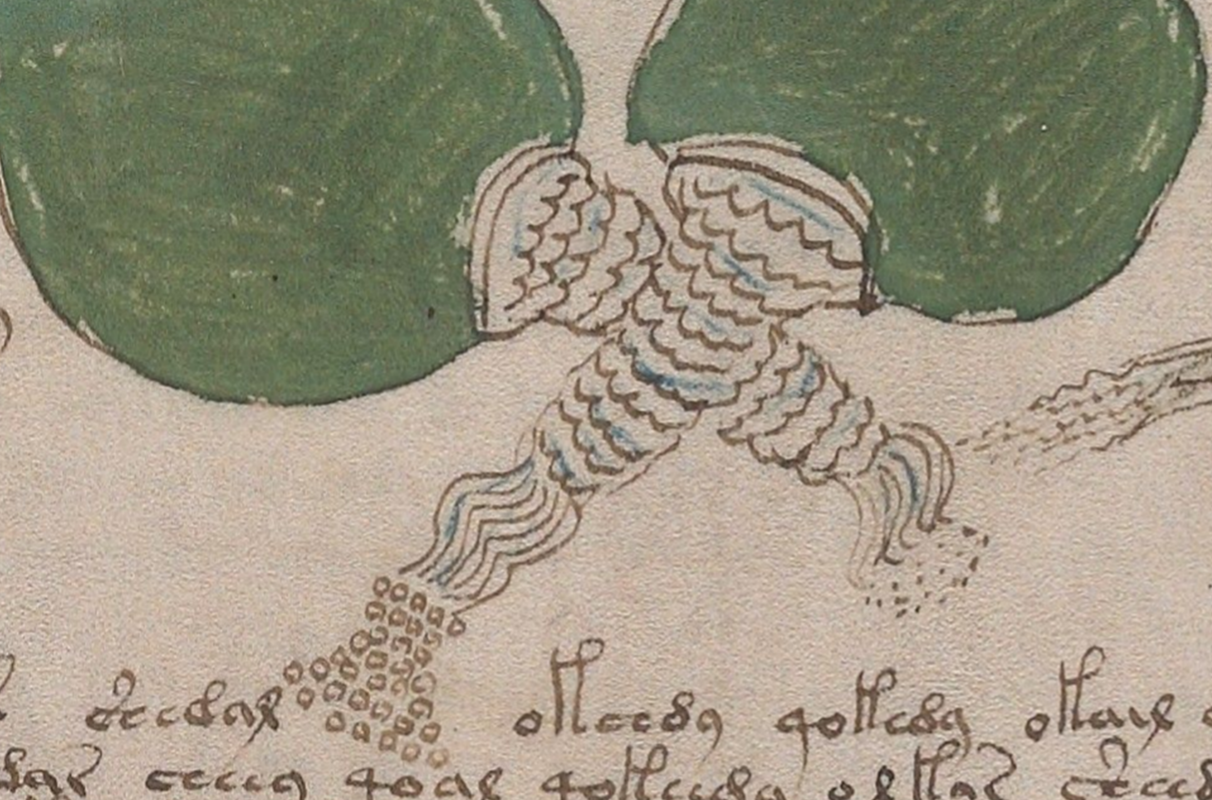
hormonal activity is taking place – and that we are witnessing one of the most energetic and transformative moments of the cycle: ovulation. From each ovary extends a whitish protrusion toward the centre – not like tentacles, but as a depiction of the very moment of release when the egg is expelled. In biological terms, this is the rupture of the follicle, where the egg – together with follicular fluid and hormones – is expelled and then captured by the fimbriae of the fallopian tube. It is this precise moment that the image captures with great accuracy.
It is important to note that the ovaries do not normally look like this. Only in the days around ovulation (days 10–11) do they take on this special structure and activity. For the rest of the month, they are more quiescent and rounded. The protrusions we see here show an active moment when the surface of the ovary is breaking.
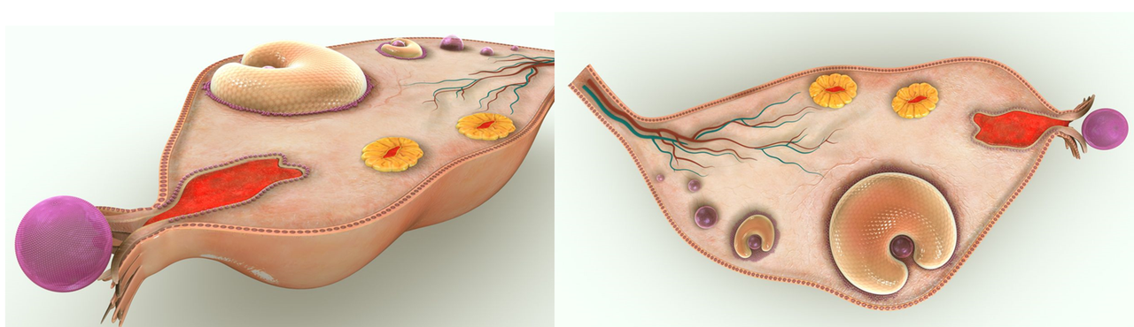
Two modern medical visualisations of the moment of ovulation. In both images, the egg (purple) is shown as just being released from the ovary, but not yet captured by the fallopian tube. The small “protrusion” on the surface shows the rupture of the follicle – the same moment also depicted in the Voynich manuscript’s image 83v with a white outflow and small droplets. The images confirm that the manuscript portrays a precise biological moment in the woman’s cycle.
The left protrusion clearly releases a series of small, round droplets or spheres, flowing out in a pattern. This illustrates that it is this side from which the egg is released, while the opposite side likely remains inactive. This corresponds to reality, where the ovaries alternate in releasing an egg from month to month – though both are hormonally influenced.
The round droplets in the image may represent:
• Follicular fluid (hormone-rich fluid that assists the egg’s transport)
• Protective layers and cells (corona radiata, zona pellucida)
• Or symbolic life energy that the illustrator has added with poetic freedom
Both protrusions form an X, which may symbolise the monthly alternation between the right and left ovary – a type of rhythmic cross-movement that gives structure and dynamism to the woman’s cycle.
The egg is then transported through the fallopian tube over the next 4–5 days, reaching the uterus around day 14–15 of the cycle – in accordance with modern biological knowledge and the medical reference image presented earlier.
Interpretation of the Woman with the Crown (folio 80v) – Reception in the Uterus
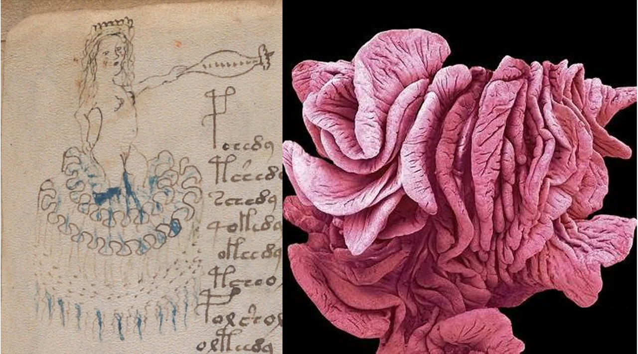
On folio 80v of the Voynich manuscript’s biological section, a woman is depicted frontally, with raised hands and a focused, almost meditative expression. Her head is adorned with a kind of crown – or rather a rhythmic, domed structure – composed of 6–7 curved segments. These arches resemble, in both form and number, the ovarian follicles before ovulation, and can be interpreted as a depiction of cyclical maturation. However, the essence of the motif does not unfold in the ovary – it unfolds in the uterus.
The woman reaches towards a long, rod-like structure with a flower-shaped opening at one end and a series of small dots along its shaft. It is natural to interpret this as a symbolic representation of the uterine lining (the endometrium) – in the phase when the body is hormonally prepared to receive a fertilized egg. The small dots may mark hormonal impulses, energy fields, or the inherent information of the egg – perhaps even a poetic allusion to genetic code. This is not anatomical precision, but a rhythmic and functional depiction of life’s gateway.
The gesture is not one of giving, but of receiving. Her hands are raised and open, as if to welcome something. The body – here visualized as a woman – receives not merely an egg, but a principle. A nascent, potential life.
The motif on 80v corresponds remarkably with the structures seen in modern microscopic images of the endometrium in the secretory phase. Here, folded tissue zones, deep recesses, and soft glandular structures can be seen – precisely the features that enable the lining to receive and hold an egg. The comparison supports that the illustrator had understanding of – or intuition about – the uterus’s receptive function and its rhythmic architecture.
This image therefore does not simply show a figure, but a symbolic and functional scene: the woman as the personification of the uterus – receptive, rhythmic, and connected to a greater cyclical movement. She does not receive randomly. She knows when she is ready.
For six centuries, the world has read the Voynich manuscript the wrong way.
Cryptographers chased letters. Scholars clung to alphabets. Mystics interpreted symbols. But they saw only shadows – and mistook them for reality.
The truth was in front of their eyes all along.
What you see in this document is not a theory – it is proof. Not an interpretation, but a pattern of mathematical precision. Not speculation, but structures repeating with surgical accuracy, page after page.
The images ARE the code. The text is only an echo.
Once you see it, the entire mystery collapses. Six hundred years of confusion vanish in a moment. The Voynich shifts from insoluble riddle to self-evident truth – so clear, so undeniable, that the only question left is: how could everyone have missed it?
This is not a hypothesis.
It is the key – and it cannot be disproven.
Excerpts from the Voynich Manuscript – The Woman’s Cycle in Encoded Form
This document presents a series of selected observations and visual excerpts from the Voynich Manuscript – a mysterious and still untranslated work from the 15th century that has baffled researchers for over a hundred years.
Rather than undertaking long, time-consuming analyses of individual pages – which can often take up to 100 hours per image – I have gathered here some of the clearest and most unmistakable signs, symbols, and structures that reveal what the manuscript is truly about: the woman’s cycle, ovulation, fertilization, hormones, and the body’s inner rhythm.
Each excerpt has been chosen because it contains distinct traces of biological or functional processes. These may include visualizations of the fallopian tube, cellular structures, menstrual rhythms, or the interrelated roles of women within a cyclical system.
The document serves as a kind of visual compendium or key extract – not as a finished analysis, but as a direct pathway into the core of the manuscript. The aim is to offer the reader an immediate view of what is otherwise often obscured by cryptic language and overextended interpretations.
I hope these excerpts can serve as a compelling introduction for new readers, while also giving experienced Voynich researchers concrete insight into the female and biological structures that form the manuscript’s underlying framework.
Analysis of the Blood Vessel in the Voynich Manuscript
The image above shows a detailed and technical construction that, in all likelihood, depicts an artery. The structure is built up in several concentric layers, precisely as we know it from modern anatomical illustrations. The outer zone is deep red, corresponding to the tunica externa, while the middle zone, with its smooth and clearly defined texture, can be interpreted as the tunica media – the muscular layer that regulates blood flow. At the top, there is a separate section with dots and patterns, which likely represents the tunica intima and the active blood flow.
This composition shows that the author of the Voynich Manuscript not only understood the structure of blood vessels but also sought to document their role in the body’s cyclical and hormonal functions. It strengthens the overall interpretation of the manuscript as a physiological and botanical guide to a woman’s inner balance.
The fact that this particular artery is depicted with such great precision – and flanked by herbal drawings on the sides – indicates that the condition of the arteries was considered a central aspect of women’s health. It is possible that plants on the nearby pages of the manuscript were intended as treatment or regulation of blood quality, circulatory rhythm, or hormone transport.
Arteries carry oxygen, hormones, and signaling substances. In connection with the woman’s cycle, they transport, among other things:
• estrogen and progesterone,
• LH and FSH (which regulate ovulation),
• nutrients and immune-active substances.
This makes the blood vessels a critical infrastructure in cycle regulation. That they are shown in detail in the manuscript must therefore be understood as a direct acknowledgment of their significance.
The image’s tubular form, layering, and color coding match almost identically with modern medical illustrations, making it extremely unlikely to be coincidental. Rather, it must be understood as part of a systematic and targeted attempt to map a woman’s internal anatomy and its rhythmic interaction with nature’s plants and patterns.
Past Misinterpretations and the Breakthrough in Understanding
It is remarkable that an image with such a clearly anatomical structure has never previously been correctly identified as an artery. Many established Voynich researchers have instead suggested that the figure depicts a strange vessel, a herb press, an alchemical instrument, or some type of plant stem. These interpretations reveal how far research into the manuscript has been from identifying the actual biological elements.
But what we see here is not symbolism and not alchemy. It is medical structure. An artery with multiple layers, precisely as it appears in modern anatomy – and even with color coding and details showing:
• inner endothelial layer,
• smooth muscles in the middle,
• outer protective layer and tissue structure.
It is simply impossible for this construction to be the result of chance. And thus, the image shows not only a deep understanding of the body’s anatomy but also that the author had a purpose:
To show how the body’s infrastructure – including the blood vessels – is connected to the woman’s cycle and the plants that influence it.
This makes the image a decisive breakthrough in Voynich interpretation. For it confirms that the manuscript is not nonsense, not a code, but an advanced anatomical and hormonal manual that must have been based on empirical knowledge and observation – long before modern medicine achieved this understanding.
Analysis of the Vein Structure – An Anatomical Breakthrough in the Voynich Manuscript
The image above most likely shows a cross-section of a vein – a motif that many have overlooked or misunderstood as some kind of container, alchemical furnace, or decorative column. But when compared with modern medical illustrations of vein structure, it becomes clear that what we see here is a fully anatomically correct depiction – complete with vein walls, layering, and the characteristic venous valves.
Veins differ markedly from arteries in several ways:
• They have thinner walls, as blood pressure is lower.
• They contain valves that prevent blood from flowing backward.
• They are often closely linked to hormonal and circulatory changes, especially in women.
The image reproduces precisely these features:
• The lower arched structures resemble the folded venous valves with almost surgical accuracy.
• The divided middle section, with distinct layers and deep grooves, corresponds exactly to how modern anatomy illustrates the tunica media and tunica intima.
• The upper part of the cylinder shows a thin, lighter layer with dots, matching the elastic inner surface where the endothelial layer lies.
The comparison between the manuscript image and the modern vein illustration shows not only a similarity in form, but layer-by-layer identity. This demonstrates with overwhelming clarity that the author of the Voynich Manuscript had detailed knowledge of vein anatomy – long before the invention of the microscope.
Function and Context in the Manuscript
It is no coincidence that there are multiple images of both blood vessels and veins in the manuscript. Their structure and condition change markedly throughout the woman’s cycle – depending on hormonal balance, blood pressure, diet, and physical condition. It is well known that estrogen and progesterone affect the elasticity, permeability, and flow of veins, and many women experience symptoms such as varicose veins, swelling, or a feeling of heaviness in the legs during certain phases of the cycle.
The manuscript’s linking of veins and plants (often placed close to each other) therefore suggests a deep understanding of how natural remedies influence specific parts of the circulatory system – not generally, but targeted to the woman’s phases and physiology.
It thus becomes increasingly clear that the Voynich Manuscript is a biological textbook for women – and not merely a collection of mysterious figures. The systematic focus on the circulatory system (arteries, veins, and later the fallopian tube) shows that the body’s transport system – and thus hormone distribution, nutrition, and reproduction – is at the center.
This image is clear evidence of a science that united plant knowledge and anatomy with cyclical understanding – and which modern medicine only caught up with much later.
Analysis of the Fallopian Tube in the Voynich Manuscript – Cyclical Variation and Precise Day Interpretation
In this section, we analyze the fallopian tube organ, as it is depicted in the Voynich Manuscript. The modern medical reference image clearly shows the organ’s structure: a tubular structure with three distinct layers – mucosa (a folded mucous membrane), muscularis (smooth muscle), and serosa (an outer connective tissue layer containing blood vessels and nerves). This anatomical structure corresponds remarkably well with the two selected Voynich illustrations, which seem to show the structure of the fallopian tube – both at rest and in function.
The fallopian tube is where the egg travels from the ovary toward the uterus. And it is precisely in this movement that the manuscript’s imagery becomes especially detailed and ingenious. The two manuscript images show different phases or stages in this process. Both figures are composed of coloured sections in cylindrical form, with characteristic “grasping arms” at the basal end. These extensions likely correspond to the fimbriae of the fallopian tube, which actively capture the egg after ovulation.
The image on the right is particularly remarkable: it shows a green basal segment with finger-like arms, a clearly marked red section in the middle, and a white top-point structure – which likely symbolises the fertilised or newly released egg. The green area at the basal end indicates an active energy phase, where the egg has just been captured. Meanwhile, the upper part reveals an open and undulating form, which can be interpreted as the movements of the mucosa layer – the folds guiding the egg forward.
This woman – depicted in the margin of the corresponding image – holds a spherical object in her hand, as if she reveres it as an offering or revelation. The symbolism is powerful: she represents the second day within the fallopian tube’s active phase, approximately day 12 of the woman’s cycle. On this day, the egg is released from the ovary and captured by the fallopian tube, which now attempts to transport it toward the uterus. Her blue and white clothing supports this image of purity and hormonal activity. The crown or pointed tip of the object she carries matches exactly the pointed form that the fallopian tube in Image 1 displays at precisely this stage of the cycle – showing that the manuscript uses colours, shapes, and “crowns” to indicate exactly which day of the cycle is depicted.
It is important to note that these images are not arbitrary variations. The fallopian tube is not static – it changes shape and appearance depending on the stage of the woman’s cycle. The many versions of this structure in the Voynich Manuscript bear witness to detailed, day-by-day knowledge of the woman’s cycle. Each “vessel” or “column” has a unique tip, a unique basal element, and a colour code that precisely indicates the point in the cycle.
This precision is extraordinary. First, because one could never map these variations in the anatomy of a deceased woman. The dynamics and structure of the fallopian tube can only be understood through living observation – requiring repeated, cycle-based examinations. This attests to an unusually high level of knowledge, likely based on experiments, observation, and experience through generations.
The manuscript’s imagery therefore seems to reflect a system in which the woman’s cycle is mapped in detail – not only in terms of internal processes (such as ovulation and hormone production), but also in how the body’s organs change in appearance and function from day to day. This cyclical rhythm is likely connected to the many plants depicted outside the structures in the manuscript. It suggests a possible relationship between hormonal balance and diet – and knowledge of how specific herbs and plants could be used to support the fallopian tube and ovulation at specific times.
All of this points to inherited, practically applied knowledge of female fertility – where each day has its signs and each organ its cyclical transformation.
A preliminary and precise proposal for image 85v/86r – The egg’s journey from ovulation to possible implantation
This is not a full analysis, but a short and focused excerpt from image 86r in the Voynich manuscript. Based on the rhythm, colours, structures, and placement of the image, it is assessed that the motif shows the egg’s journey from ovulation through the fallopian tube and leading to either implantation in the uterus – or rejection. The interpretation is based on visual patterns and symbolism and should be read as a qualified proposal within a broader overall understanding of the manuscript’s biological sections.
The image contains:
• 6 towers (functional phases in the fallopian tube)
• 5 inner rings (the cilia of the fallopian tube and hormonal rhythms)
• 6 outer rings (stages of the uterus)
• 4 outer points (possible outcomes for the egg)
• An overarching “matrix” (hormonal network)
• Names, eggs, dots, and rhythmic repetitions
1. The central field: The 6 towers
The towers in the centre are not decorative but precise visualisations of the fallopian tube’s phases. They represent functional phases, not time units. Each tower form differs anatomically and rhythmically.
At the top are seen spiral-shaped drops (blue), pointing to the eggs just released being “charged” – the same symbol appears in spiral image 156 as an ovulation marker.
2. The inner zone: 5 cilia rings = the fallopian tube
Around the towers, five concentric rings are seen, filled with eggs and cilia-like structures. The eggs vary in dots and colour, which is interpreted as different hormonal and cellular stages.
• 1st ring: Active collection – dense cilia, eggs just released
• 2nd ring: Colour shift – oestrogen stimulates movement
• 3rd ring: Dot patterns = beginning cell division
• 4th ring: Sequence of eggs with and without dots, initial selection
• 5th ring: Oestrogen level declines, transition to the outer rings
The pattern starts at the bottom (at 6 o’clock) and follows clockwise – the rhythm is precise, and the style shows a clear sequential progression.
3. The outer zone: 6 uterine rings
Around the five cilia rings, six outer rings are shown, each with eggs in different stages.
• The eggs are now without blue colour = oestrogen level declines, progesterone takes over
• The cilia are still shown but are now interpreted as a symbol of energetic flow
• The eggs are distributed in shifting densities = variation in hormonal receptivity of the uterus
Some rings show eggs in close symmetry – others show asymmetry = biological uncertainty.
4. The hormonal “garment”: The matrix network above the towers
Above the 6 towers hovers a network of stars, drops, and blue lines – this network is interpreted as a hormonal matrix that integrates all functions.
The matrix functions as:
• Coordinated hormonal pulse (oestrogen → progesterone)
• Coordination between cilia, secretion, and energy
• Visual and rhythmic coupling to spiral image 156
The net structure shows that the cycle is not divided but coherent and rhythmic all the way.
5. The 4 outcome scenarios at the image’s periphery
Four small, distinctive patterns at the image’s periphery show possible endpoints for the egg’s journey.
These scenarios confirm that the artist understood the beginning of life as conditional and multi-phased.
6. Symbols, names, and energies
Below the towers are seen names – not as proper names, but as energetic signatures for each functional phase. The writing style does not differ from the rest of the manuscript, but the placement suggests that the names are part of the towers’ functions.
• Can be interpreted as hormonal keys
• Or as a code for functional or rhythmic energy
• Likely connection to interpretations in the spiral image
7. Visual structure and symmetry
The geometric structure is not random:
• 6 towers = functional stages
• 5 rings = fallopian tube
• 6 rings = uterus
• 4 endings = biological outcomes
In total = 21 units → the same structure as a typical fertile cycle (21–35 days), indicating an awareness of cyclical rhythms.
Conclusion – Image 86r is a functional mapping
The four orange arrows show how the cycle in folio 85v/86r moves in four directions out from the central field.
The centre (with the six towers) functions as a central rhythmic and biological axis, from which the egg’s journey branches out – either in various phases of development or towards possible outcomes. The arrows mark the structure’s division into four functional paths or flows, each encompassing a distinct theme in the cycle.
This image is not decorative – it is a biological, microscopic, and rhythmic model of the whole journey from ovulation to either life or rejection.
It combines:
• Anatomy
• Biochemistry
• Symbolism
• Geometry
• Understanding of energy
It demonstrates a deep, functional understanding of a woman’s cycle, which has only been confirmed today through microscopy and reproductive biology.
Final note – The spiral’s microscopic anchoring and the egg’s rhythmic journey
This image functions as a focused zoom into the egg’s path from ovulation to implantation and connects the spiral’s macro level with the biological micro level. Where the spiral image (f.156) showed the egg’s rhythm and cycle in large format, image 86r appears as the microscopic detailed view, where the egg is followed day by day and layer by layer.
This is one of the strongest visual confirmations that the Voynich manuscript contains an advanced understanding of a woman’s reproductive cycle – seen from within – with a precision and structure which has only been confirmed today through microscopy and modern reproductive biology.
The image depicts not merely a biological sequence, but a complete cyclical and stage-by-stage description of the egg’s journey:
• From hormonal activation in the ovaries
• Through the fallopian tube and the cilia’s rhythmic cooperation
• And further to the uterus, where it either implants or is rejected
Each part is shown with:
• Colour coding (blue, green, white)
• Microscopic motifs (eggs with dots, cilia, drops)
• And a geometric, rhythmic structure (towers, rings, matrix)
The image is both physical and symbolic. It is a mapping of the woman’s inner rhythm and fertility – not merely as a biological system, but as an energetic and consciousness-based process, where all layers are connected in one living whole.
The symbolism of the cilia – the inner flow
Even though the uterus anatomically does not contain cilia like the fallopian tube, the image’s outermost ring still shows cilia-like patterns. This should be understood symbolically – as a sign of the egg’s continued movement and inner advance toward the correct implantation site.
The egg typically seeks toward the lower and rear parts of the uterine lining, and precisely this search and rhythmic passage is here illustrated as a visual extension of the fallopian tube’s flow. The cilia should not be read as anatomical elements, but as energetic bearers of life’s pulse.
The Woman’s Contact with the Stream of Life – An Energetic Narrative of Ovulation and Fertile Activation (Voynich f83v)
When one first observes the image on folio 83v of the Voynich manuscript – the naked woman in a vessel, framed by a green arch and holding blue substance in her hands – it may appear to be a mythical or decorative motif. Yet beneath its simple exterior lies a deeply complex narrative about the beginning of life, the woman’s cyclical biology, and the precise moment when fertility is activated.
The woman is literally standing inside the uterus – the illustrated vessel is not arbitrary but anatomically and symbolically identical to the womb. The green arch above her is not mere ornamentation; it represents an aesthetic and functional rendering of an artery – one of the major blood vessels supplying the uterus and ovaries. Through this channel, the life-giving signaling substances (hormones) and nutrients arrive.
We see the woman raising her hand to receive something blue from the green tube. The blue substance continues its journey down into the vessel – and further down through a vertical tube decorated with small marks and energy lines. This tube is especially remarkable: it shows that only one vital pathway is active – exactly as in reality, where only one fallopian tube at a time transports an egg toward the uterus.
The blue substance may be interpreted as the egg itself, or perhaps as the hormonal impulse – in particular, the luteinizing hormone (LH), which is released once in each cycle and is essential for ovulation to occur. It is precisely timed and comes in a short, powerful surge – perfectly matching the blue energy flowing down through the woman’s body.
The choice of a green tube is not coincidental. In many cultures and symbolic systems, green represents life, energy, growth, and fertility. In this context, it is natural to see the green arch as a stream of life energy – a channel activated once per cycle, carrying the potential for fertility. One can imagine that the authors of the manuscript sought to show that the woman, in this moment, is not merely a passive recipient but a conscious participant in life’s circulation. Her hand, reaching up to receive the impulse, demonstrates contact, awareness, and participation in the body’s deepest rhythms.
Further down, we see a fimbriated tube – precisely one channel, decorated with small lines and symbols. This tube corresponds to the fallopian tube – and here one of the manuscript’s most remarkable details is emphasised: there are not two tubes depicted. Only one. For this is how the body functions – only one fallopian tube is active in each cycle. This is a biological fact that only became commonly known through modern anatomy. That the Voynich manuscript depicts it with such accuracy suggests it rests upon detailed, practical knowledge of the woman’s inner functions.
When compared with modern anatomical images of the blood supply to the uterus, ovaries, and fallopian tube (as shown above), we see how every colour, movement, and body part in the Voynich image corresponds to something real – and how, at the same time, it is elevated into a symbolic narrative of life, energy, and timing.
This image is therefore not merely an illustration of anatomy – it is a profound energetic mapping of the woman’s fertile moment. The green arch is not just blood – it is the life-stream. The blue substance is not merely liquid – it is hormone, egg, impulse. And the woman is not passive – she is both portal and participant in life’s eternal movement.
Note on Symbolism
It is important to understand that the woman in the image should not necessarily be perceived as a physical person in an anatomical sense. Instead, she functions as a symbol of the female cycle and the inner energetic process. When she stands “inside the uterus,” it is not because we are seeing literal anatomy, but because the manuscript employs a visual and sensory language to explain the body’s own mechanisms from within.
That the woman receives the blue substance with her hand and lets it flow onward is therefore not merely a bodily gesture – but a metaphor for conscious contact with the hormonal signal or the energetic flow that activates fertility. Everything in the image must be read with this symbolic layer in mind – something that, incidentally, applies to the entire Voynich manuscript.
It is also important in this context that the woman stands naked. This is not accidental. In the Voynich manuscript, nakedness functions as a symbol of biological vulnerability, openness, and activation – it marks that something fundamental is occurring in the body’s circulation, and that the woman is here in one of the most vital phases of the monthly cycle. She is not a passive observer – she is herself the organism in which it occurs, and the blue substance and green connections are not without significance.
Analysis of Image 83v – The Ovaries in Function During Ovulation
In this image from the Voynich manuscript (folio 83v), two large, dark green spheres can be seen, which can with great confidence be interpreted as the woman’s ovaries. The drawing style is bold and focused, and the colour choice itself is a clear signal: the two spheres are painted in a distinctive deep green tone, distinctly different from the surrounding structures. This indicates that intense
hormonal activity is taking place – and that we are witnessing one of the most energetic and transformative moments of the cycle: ovulation. From each ovary extends a whitish protrusion toward the centre – not like tentacles, but as a depiction of the very moment of release when the egg is expelled. In biological terms, this is the rupture of the follicle, where the egg – together with follicular fluid and hormones – is expelled and then captured by the fimbriae of the fallopian tube. It is this precise moment that the image captures with great accuracy.
It is important to note that the ovaries do not normally look like this. Only in the days around ovulation (days 10–11) do they take on this special structure and activity. For the rest of the month, they are more quiescent and rounded. The protrusions we see here show an active moment when the surface of the ovary is breaking.
Two modern medical visualisations of the moment of ovulation. In both images, the egg (purple) is shown as just being released from the ovary, but not yet captured by the fallopian tube. The small “protrusion” on the surface shows the rupture of the follicle – the same moment also depicted in the Voynich manuscript’s image 83v with a white outflow and small droplets. The images confirm that the manuscript portrays a precise biological moment in the woman’s cycle.
The left protrusion clearly releases a series of small, round droplets or spheres, flowing out in a pattern. This illustrates that it is this side from which the egg is released, while the opposite side likely remains inactive. This corresponds to reality, where the ovaries alternate in releasing an egg from month to month – though both are hormonally influenced.
The round droplets in the image may represent:
• Follicular fluid (hormone-rich fluid that assists the egg’s transport)
• Protective layers and cells (corona radiata, zona pellucida)
• Or symbolic life energy that the illustrator has added with poetic freedom
Both protrusions form an X, which may symbolise the monthly alternation between the right and left ovary – a type of rhythmic cross-movement that gives structure and dynamism to the woman’s cycle.
The egg is then transported through the fallopian tube over the next 4–5 days, reaching the uterus around day 14–15 of the cycle – in accordance with modern biological knowledge and the medical reference image presented earlier.
Interpretation of the Woman with the Crown (folio 80v) – Reception in the Uterus
On folio 80v of the Voynich manuscript’s biological section, a woman is depicted frontally, with raised hands and a focused, almost meditative expression. Her head is adorned with a kind of crown – or rather a rhythmic, domed structure – composed of 6–7 curved segments. These arches resemble, in both form and number, the ovarian follicles before ovulation, and can be interpreted as a depiction of cyclical maturation. However, the essence of the motif does not unfold in the ovary – it unfolds in the uterus.
The woman reaches towards a long, rod-like structure with a flower-shaped opening at one end and a series of small dots along its shaft. It is natural to interpret this as a symbolic representation of the uterine lining (the endometrium) – in the phase when the body is hormonally prepared to receive a fertilized egg. The small dots may mark hormonal impulses, energy fields, or the inherent information of the egg – perhaps even a poetic allusion to genetic code. This is not anatomical precision, but a rhythmic and functional depiction of life’s gateway.
The gesture is not one of giving, but of receiving. Her hands are raised and open, as if to welcome something. The body – here visualized as a woman – receives not merely an egg, but a principle. A nascent, potential life.
The motif on 80v corresponds remarkably with the structures seen in modern microscopic images of the endometrium in the secretory phase. Here, folded tissue zones, deep recesses, and soft glandular structures can be seen – precisely the features that enable the lining to receive and hold an egg. The comparison supports that the illustrator had understanding of – or intuition about – the uterus’s receptive function and its rhythmic architecture.
This image therefore does not simply show a figure, but a symbolic and functional scene: the woman as the personification of the uterus – receptive, rhythmic, and connected to a greater cyclical movement. She does not receive randomly. She knows when she is ready.